Schistosoma mansoni, blood fluke
Schistosoma mansoni is digenic trematode ("digenic" means that its lifecycle includes two hosts - definitive and intermediate) of the superfamily Schistosomatoidea.
Intermediate host of S. mansoni are snails of the genus Biomphalaria (Planorbidae family).
S. mansoni occurs in Africa, Madagascar, parts of South America (such as Venezuela and Brazil), Puerto Rico and the West Indies.
S. mansoni is found in rodents and primates but primary target of the infection are humans.
Back to top Nemose
Life cycle of Schistosoma mansoni
egg
female discharges up to 300 eggs per day singly (not in batches as Schistosoma japonicum); into mesenteric venule of definitive host (human); the eggs eventually obstruct blood flow in the venule causing a partial necrosis in the intestinal wall; hence, the eggs are dropped of into the lumen of the intestine, and passed to feces; eggs measure ~140 X 60 µm in diameter (bigger than eggs of Schistosoma japonicum)
miracidium
a free-swimming larva; once the egg is released into environment, miracidium hatches immediately and starts swimming; if it happened to swim into a snail of species Biomphalaria sp., it enters the snail and starts its first life as a parasite
sporocyst
a sac-like secondary larval stage; miracidium transforms into a primary (mother) sporocyst; germ cells within the primary sporocyst begin dividing to produce secondary (daughter) sporocysts, which migrate to the snail hepatopancreas; once at the hepatopancreas, germ cells within the secondary sporocyst begin to divide again, this time producing thousands of new parasites, known as cercariae, which are the larvae capable of infecting mammals
cercarium
an infectious form of Schistosoma which infects their hosts by direct skin penetration; cercariae emerge daily from the snail host; they are highly motile, alternating between vigorous upward movement and sinking; cercarium attaches to the human skin and secretes proteolytic enzymes helping it to enter into cuteneous capillary vessel; upon the penetration the cercarium sheds its tail and transforms into schistosomulum
schistosomulum
a tailless cercarium; after penetration and spending a few days in the skin, schistosomula migrate to the lungs (in 3-4 days), and after passing through the pulmonary capillaries, enter the systemic circulation and, eventually, are carried to the mesenteric vein of the host; there they mature into adult schistosomes in a month; female schistosomes do not maturate without a mature male and females schistosomes from single sex infections are underdeveloped and exhibit immature reproductive system; male schistosome holds female between its gynecophoral canal; at this time, the infection of the final host is complete and sexual reproduction of the parasite begins
adult
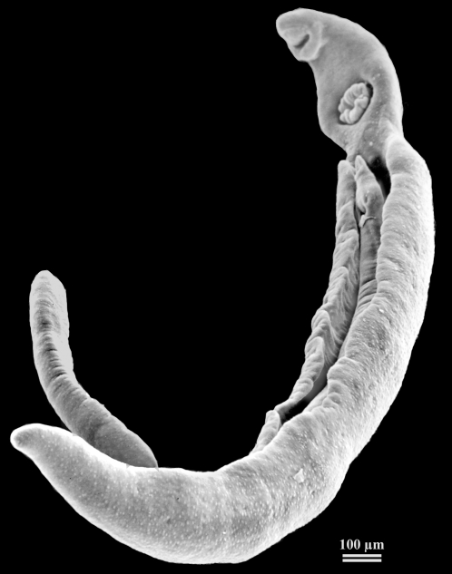
adult males are approximately 1 cm long and 0.11 cm wide; adult females are approximately 1.4 cm long and 0.016 cm wide; the adults can live for years; male and female are always hugged together; up to half the eggs released by the worm pairs become trapped in the mesenteric veins, or will be washed back into the liver, where they will become lodged; trapped eggs mature normally, secreting antigens that elicit a vigorous immune response
References
Brindley PJ, Mitreva M, Ghedin E, Lustigman S. Helminth genomics: The implications for human health. PLoS Negl Trop Dis. 2009 Oct 26;3(10):e538.

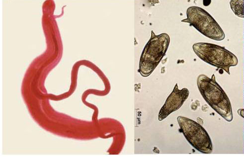
(Left) A pair of adult worms of the blood fluke Schistosoma mansoni; the more slender female worm resides in the gynecophoral canal of the thicker male. The worms are about 1.5 cm in length, and live for many years .
(Right) Eggs of Schistosoma mansoni. The egg is about 150×50 µm in dimension; the lateral spine is diagnostic for S. mansoni in comparison to the other human schistosome species. Fibrotic responses to schistosome eggs trapped in the intestines, liver, and other organs of the infected person are the cause of the schistosomiasis pathology and morbidity.

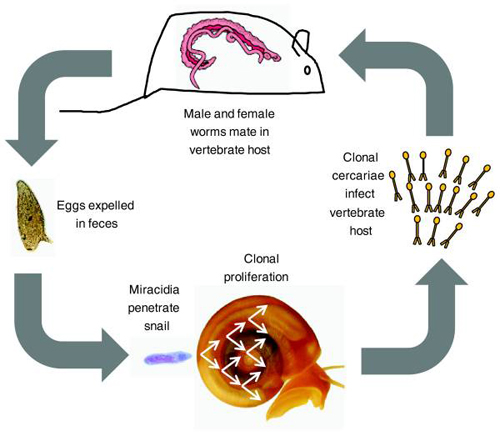
Schistosoma mansoni life cycle.
The life cycle involves both an aquatic snail intermediate (Biomphalaria spp.) and a human definitive host. Mice and hamsters can be used to maintain the life cycle in the laboratory. Male (broad pink and red) and female (skinny pink) adult worms are found in the venules draining the intestine. Eggs pass through the intestine and out of the body with the feces. The eggs hatch in fresh water, and motile miracidia actively search for snails. Following penetration into the snail host, miracidia differentiate into sporocysts. Sporocysts proliferate asexually in the snail, eventually releasing motile clonal cercariae into the water. Cercariae penetrate the unbroken skin of a mammalian host, and then migrate through the bloodstream to the hepatic portal system where they develop into adults. In the laboratory, the entire life cycle takes 75 to 90 days to complete. S. mansoni is a conventional dioecious diploid, except for the fact that larval forms replicate asexually within the snail intermediate host. This aids in the staging of genetic crosses because clonally generated male and female larvae from different snails can be used to infect mice.
Beltran S, Cézilly F, Boissier J. Genetic dissimilarity between mates, but not male heterozygosity, influences divorce in schistosomes. PLoS One. 2008 Oct 8;3(10):e3328.
